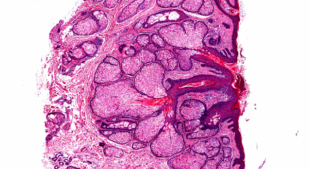
Answer of Dermatopathology Case 30
Xanthelasma
Visit: Xanthelasma;
Visit: Dermatopathology site;
Visit: Verruciform Xanthoma ;
Visit: Gastric Xanthoma
Visit: Eye Pathology Online .
Abstract:
Xanthelasma and juvenile xanthogranuloma in a 7-year-old boy. Ann Dermatol Venereol. 2009 Oct;136(10):723-6. Epub 2009 May 15.
BACKGROUND: Palpebrum xanthelasma is the most common type of xanthoma seen inadults but it is extremely rare in children. We report an original case of bilateral xanthelasma palpebrarum associated with juvenile xanthogranuloma (JXG) in a 7-year-old child. Only two cases of xanthelasma in children have been described to date. The association of xanthelasma and JXG has never been described. PATIENTS AND METHODS: A 7-year-old boy presented xanthelasmas on both eyelids. At the same time, pinkish JXG papules appeared on the child's trunk. The boy had been diagnosed at the age of 10 months with myelogenous leukaemia, which was in remission. He also had a familial history of hypercholesterolaemia. The skin lesions were removed and microscopic examination confirmed the diagnosis of xanthelasmas and JXG. DISCUSSION: This patient's presentation is unusual inseveral respects: the presence of xanthelasma in a child, appearance of JXG at an advanced age, and the association of these two diseases in a child with a past history of leukaemia. The occurrence of these skin lesions did not appear to be linked to the history of malignant blood disease in this patient.
Unknown: bilateral symmetrical papules on the eyelid.Dermatol Online J. 2009 May 15;15(5):13.
Eyelid lesions frequently are a diagnostic challenge. We report a case of a 46-year-old woman with a 5-year history of yellowish symmetric progressivelygrowing papules on the eyelids, resembling xanthelasma. A skin biopsy wasperformed that revealed the rare variant of clear cell syringoma. The lesionswere treated with CO2 laser and surgical excision; there was no evidence of recurrence after 6 months of follow-up.
Xanthelasma palpebrarum and its relation to atherosclerotic risk factors and lipoprotein. Int J Dermatol. 2008 Aug;47(8):785-9.
OBJECTIVES: To investigate the association between xanthelasma, atherosclerotic risk factors, and lipoprotein (Lp) (a), and to determine whether xanthelasma may be a cutaneous marker for atherosclerosis. METHODS: One hundred consecutive patients with xanthelasma and 100 age- and sex-matched patients without xanthelasma, seen during the same time period (controls), were included in this study. The prevalence of cardiac risk factors, the rates of atherosclerotic disease, Framingham risk scores, and Lp (a) levels were compared between the patient groups. RESULTS: Hyperlipidemia was found to be significantly more common in patients with xanthelasma (P = 0.001) ; however, the rate of clinically overt cardiovascular disease and future cardiovascular risk, assessed by the Framingham risk score, were similar between the groups. No significant difference was observed in serum Lp (a) levels between the groups. CONCLUSIONS: In patients with xanthelasma, no increase was observed in the rate or risk of cardiovascular disease. Moreover, no relationship was found between Lp (a) levels and xanthelasma.
Xanthelasma and lipoma in Leonardo da Vinci's Mona Lisa.Isr Med Assoc J. 2004 Aug;6(8):505-6.
The painting Mona Lisa in the Louvre, Paris, by Leonardo da Vinci (1503-1506), shows skin alterations at the inner end of the left upper eyelid similar to xanthelasma, and a swelling of the dorsum of the right hand suggestive of asubcutaneous lipoma. These findings in a 25-30 year old woman, who died at the age of 37, may be indicative of essential hyperlipidemia, a strong risk factor for ischemic heart disease in middle age. As far as is known, this portrait of Mona Lisa painted in 1506 is the first evidence that xanthelasma and lipoma were prevalent in the sixteenth century, long before the first description by Addison and Gall in 1851.


Comments
Post a Comment