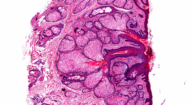
Answer of Dermatopathology Case 60
Spitz Nevus
Visit: Dermatopathology site
Visit: Pathology of Spitz Nevus
The spitz nevus: review and update.Clin Plast Surg.2010 Jan;37(1):21-33.
The Spitz nevus is a relatively common skin lesion in children and is less commonly seen in adults. The lesion is defined by the presence of distinctive-appearing spindle or epithelioid cells on light microscopy in a recognizable nevus-like pattern. Spitz lesions share features with melanoma on light microscopic examination. When Spitz features are atypical or typical features are absent, distinction from melanoma can be difficult. A spectrum of pathology of Spitz lesions can be found from lesions that are benign and typical to lesions that are atypical with melanoma-like features and frank melanoma. There is significant interobserver variation in interpretation of Spitz lesions. The lack of uniformly applied criteria for distinction of light microscopic grades and the confusion in diagnostic terminology demonstrate the difficulty in the pathologic interpretation of these lesions. Exciting progress has been made recently in ancillary testing that will likely be helpful in determining in more detail the biologic nature of these lesions, in better differentiating the benign Spitz lesions from malignant lesions, and in eventually improving treatment recommendations.
Pediatric atypical spitzoid neoplasms: a review with emphasis on 'red' ('spitz') tumors and 'blue' ('blitz') tumors. Dermatology.2010;220(4):306-10. Epub 2010 May 6.
BACKGROUND: The diagnosis of pediatric atypical Spitz nevus/tumors (pASNT) is an emerging challenge in clinical dermatology and dermatopathology.
OBJECTIVE AND METHODS: We review the main clinicopathologic issues raised by pASNT and describe 2 examples of different clinicopathologic subsets of lesions.
RESULTS: While Spitz/Reed nevi are commonly small- to medium-sized, tan to black plaques, pASNT are large and nodular, either 'red' (dotted and/or polymorphous vascular pattern on dermoscopy; spindle and/or epithelioid tumors on histopathology: Spitz tumors, sensu strictiori) or 'blue' (homogeneous blue color on dermoscopy; intimate admixture of epithelioid cells and heavily pigmented dendritic cells on histopathology: Blitz tumors or pigmented epithelioid melanocytomas).
CONCLUSIONS: Different clinicopathologic settings of pASNT probably exist. Dermoscopy can aid in their recognition and classification.
Spitz nevus in a Hispanic population: a clinicopathological study of 130 cases.Am J Dermatopathol.2010 May;32(3):267-75.
Spitz nevus is an uncommon melanocytic nevus distinctive by its epithelioid and spindled melanocytes. Many studies have attempted to characterize Spitz nevus, but none of them in a Hispanic population. Our aim is to characterize the clinical and histopathological presentation of the Spitz nevus in a Hispanic population. A retrospective study was carried out from our files that included those cases histopathologically diagnosed as Spitz nevus. A blinded examination was performed to evaluate the histopathological characteristics of 130 lesions. The demographics of the patients, the anatomic location, and the accuracy of the clinical diagnosis were analyzed. Eighty-one females and 49 males (ratio of 1.7:1) were included in the study. The mean age was 18.8 years. Overall, the most common location was the lower extremities (41%), followed by the upper extremities (27%), trunk (16%), and head and neck (16%). The nevi followed a similar anatomic distribution in females and males. The lesions were clinically diagnosed with accuracy in 20% of the cases and characterized as a pigmented papule in 42% of the cases. Upon histopathological evaluation, most nevi exhibited symmetry (84%), were well circumscribed (91%), and exhibited epidermal hyperplasia (69%). The junctional type was seen in 42% of the cases, the compound type in 38%, and the dermal type in 20%. Sixty-eight percent of nevi were mostly composed of epithelioid melanocytes, the spindled-shaped melanocytes predominated in 17% of cases, and 12% were composed of both epithelioid and spindled-shaped melanocytes. Multinucleated melanocytes were seen in 7% of nevi, mostly in the epithelioid Spitz nevus (67%). Abundant melanin was observed in 51 cases, from which the most common variant was the classic Spitz nevi. The typical dull eosinophilic globules (Kamino bodies) were observed in a minority of the cases (11%), mostly in the classic Spitz nevus. The most common variant was the classic Spitz nevus (65%), followed by the dermal Spitz nevus (15%). In conclusion, Spitz nevus in a Hispanic population most commonly presents as a pigmented papule on the lower extremities irrespective of sex and age. It is characterized by a melanocytic proliferation most commonly composed of nested epithelioid melanocytes in a junctional or compound arrangement, with the presence of abundant melanin.
Spitz nevus: a clinicopathological study of 349 cases. Am J Dermatopathol.2009 Apr;31(2):107-16.
Spitz nevus is an infrequent, usually acquired melanocytic nevus composed of epithelioid and/or spindle melanocytes that can occasionally be confused with melanoma. Currently, there are no immunohistochemical markers or molecular biology techniques that can be used to make an entirely safe diagnosis of Spitz nevus or melanoma in problematic cases. A retrospective study has been carried out that included all the cases diagnosed as Spitz nevus from our files. Follow-up information of the patients was unavailable. Three hundred forty-nine cases of unequivocal Spitz nevi were included, and their clinical and histopathological parameters were reviewed. One hundred and forty patients (40%) were 15 years old or younger, with a male to female ratio of 1:1. In patients older than 15 years, there was an evident predominance of women, with a male to female ratio of around 1:3. Spitz nevus was most commonly located on the lower extremities, followed by the trunk in both children and adults. Despite the fact that the head and neck were the third most common location in children, it was a much more frequent location in children than in adults. The constitution by epithelioid and/or spindled cells was the only histopathological finding present in 100% of cases. The other pathological findings studied were, from more to less frequent: maturation (72%), inflammatory infiltrate (70%), epidermal hyperplasia (66%), melanin (50%), telangiectasias (40%), Kamino bodies (34%), desmoplastic stroma (26%), mitosis (23%), pagetoid extension (13%), and hyalinization of the stroma (8%). Hyalinization was the only histopathological parameter that was statistically more frequent in adults than in children.
Spitz nevus with an uncertain malignant potential.Rom J Morphol Embryol. 2009;50(2):275-82.
We present the case of 10-year-old girl who have had from birth a plane tumor, of tan color, 3-4 mm of diameter, localized on the face on the cutaneous part of the superior lip. This tumor has been stabile until 8-year-old. Then, after repeated sunlight exposures, the lesion has become more stark, hemispheric in shape, has increased in size becoming about 5-6 mm, with irregular borders, and after an accidental traumatism it began to bleed. We have performed the electroexcision of the lesion for diagnostic and therapeutic purpose. The histopathologic exam distinguished typical images of Spitz nevus on some of the histological sections but also of melanocytary tumor with uncertain malignant potential on the others where atypical mitoses localized in the deeper component of the tumor are being noticed. The immunohistochemical assessment of the tumoral cells showed positivity for the melanocytic markers HMB45 and Melan A, within junctional intraepidermic nevic cells and in the nevic cells from superficial dermis, and also for CD44 protein (belonging to the adhesion molecules family). However, cyclin D1 was positive in rare nevic cells, and the proliferation rate of the tumor was small, with a proliferation index for Ki67 lesser than 5%. The correlation between histopathological and immunohistochemical data conducive to final diagnosis of Spitz nevus with uncertain malignant potential. The clinical evolution confirmed the histopathological diagnosis by the fact that the patient did not presented clinical signs of local recurrences or metastasis at three years after the excision of the tumor.


Comments
Post a Comment