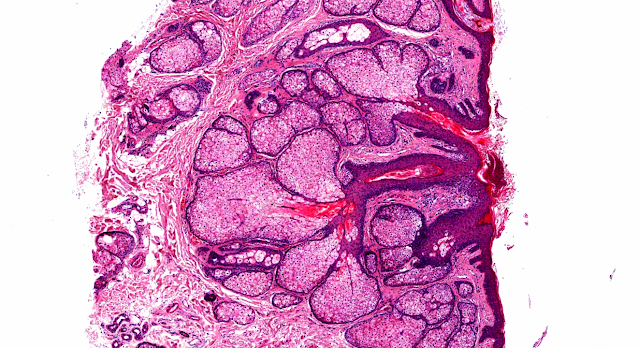
Answer of Dermatopathology Case 61
Targetoid Hemosiderotic Hemangioma
(Hobnail Hemangioma)
Visit: Dermatopathology site
Visit: Targetoid Hemosiderotic Hemangioma (Hobnail Hemangioma)
Abstract:
Hobnail hemangiomas (targetoid hemosiderotic hemangiomas) are true lymphangiomas.J Cutan Pathol.2004 May;31(5):362-7.
BACKGROUND: Hobnail hemangioma (targetoid hemosiderotic hemangioma) is a small benign vascular tumor of the superficial and mid-dermis. In contrast to its well-characterized histology, it has been unclear whether this tumor arises from blood vessel endothelial cells (BECs) or lymphatic vessel endothelial cells (LECs).
METHODS: We analyzed 10 hobnail hemangiomas by immunohistochemistry, using the recently described lymphatic endothelial cell marker, D2-40. For comparison, CD31, CD34, and alpha-smooth muscle actin expression were studied in consecutive sections of the paraffin-embedded tissues.
RESULTS: In all analyzed vessels, D2-40 labeled exclusively LECs, whereas BECs were consistently negative. In contrast to capillary BECs, either neighboring the tumors or intermingled, neoplastic endothelial cells of all 10 hobnail hemangiomas were strongly labeled by D2-40.
CONCLUSIONS: The results suggest a lymphatic origin for hobnail hemangiomas. This view is further supported by the CD34 negativity of endothelial cells and the lack of actin-labeled pericytes in hobnail hemangiomas, both characteristic of lymphatic vessels. Moreover, our analysis revealed that microshunts between neoplastic lymphatic vascular channels and small blood vessels occur, explaining some features of hobnail hemangiomas, such as aneurysmatic microstructures, erythrocytes within and beneath neoplastic vascular spaces, inflammatory changes, scarring, and interstitial hemosiderin deposits.
Hobnail hemangioma ("targetoid hemosiderotic hemangioma"): clinicopathologic and immunohistochemical analysis of 62 cases. J Cutan Pathol. 1999 Jul;26(6):279-86.
Hobnail hemangioma, also known as "targetoid hemosiderotic hemangioma", represents a distinctive, benign vascular tumor, characterized histologically by a biphasic growth pattern of dilated vascular structures in the superficial dermis lined by prominent hobnail endothelial cells, and collagen dissecting, rather narrow neoplastic vessels in deeper parts of the lesion. We analyzed the clinicopathologic and immunohistochemical features in a series of 62 cases. Patient age range was 6-72 years (median: 32 years); 34 patients were male and 25 female. Clinically, a broad variation of diagnoses ranging from hemangioma to dermal melanocytic nevus and fibrous histiocytoma was suggested. Nineteen tumors arose in the lower and 13 in the upper extremities, 12 on the back, 8 in the buttock and hip region, and one case on the chest wall. Follow-up information on 35 patients (range from 1 to 4 years; mean: 1.5 years) revealed no local recurrence nor systemic metastasis. All neoplasms were located in the dermis and showed a broad morphologic spectrum in dependence of the age of the lesions. In addition to lesions resembling cavernous lymphangioma or lymphangioma circumscriptum, neoplasms were seen with morphologic features reminiscent to retiform hemangioendothelioma, progressive lymphangioma and so-called Dabska's tumor. Immunohistochemistry performed in 28 cases showed positive staining of tumor cells for CD31 in all cases tested, whereas only 3 out of 28 cases stained completely positive for CD34. In addition 4 out of 8 cases stained positively for vascular endothelial growth factor receptor-3 (VEGFR-3). Neoplastic endothelial cells were surrounded by actin-positive pericytes in only 7 out of 27 cases tested. Hobnail hemangioma occurs more frequently in male patients and arises commonly in the extremities and the trunk. Histologic and immunohistochemcial features suggest a lymphatic line of differentiation for this distinctive vascular neoplasm.
Targetoid haemosiderotic haemangioma: dermoscopic monitoring of three cases and review of the literature.Clin Exp Dermatol.2005 Nov;30(6):672-6.
Targetoid haemosiderotic haemangioma represents a new, rarely reported, distinctive, benign vascular tumour, characterized histopathologically by a biphasic growth pattern of dilated vascular structures in the superficial dermis lined by prominent hobnail endothelial cells and collagen dissecting, rather narrow neoplastic vessels in deeper parts of the lesion. In the initial stage, the lesion is seen as a small purple or violaceous papule, 2--3 mm in diameter. Over time, the ecchymotic ring expands peripherally until it disappears spontaneously. In the later stages, however, the central papule remains as a slightly raised dermal lesion with a purple to brownish discolouration. We report three cases whose repetitive cyclic morphological changes of targetoid haemosiderotic haemangiomas were monitored dermoscopically at 3-month follow-ups. Histopathological examination of each lesion identified the features of targetoid haemosiderotic haemangioma. To the best of our knowledge, our three cases are the first reported in the literature of targetoid haemosiderotic haemangiomas that were regularly monitored by dermoscopic examinations, enabling development of the different stages of the same lesion to be followed.


Comments
Post a Comment