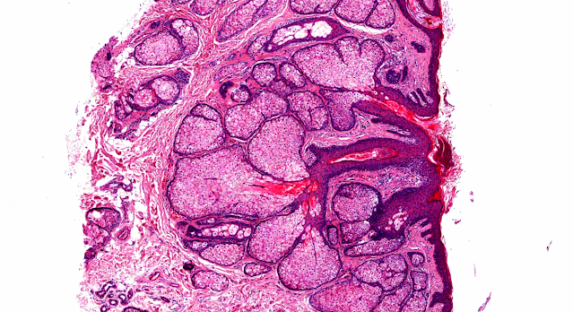
Answer of Dermatopathology Case 62
Visit: Dermatopathology site
Visit: Pathology of Chondroid Syringoma
Abstract:
Chondroid syringoma: a rare tumor of the chest wall. Ann Thorac Surg.2010 Mar;89(3):983-5.
Chondroid syringoma, an uncommon, slow-growing, benign, sweat-gland tumor located on the upper right chest wall of a 66-year-old woman is presented. This skin adenexal tumor is typically located on the head and neck region. The unusual location of chondroid syringoma made an accurate preoperative diagnosis difficult, and diagnosis was achieved only by excisional biopsy and histopathologic examination. Total surgical excision remains the best therapeutic option to avoid tumor recurrence and close follow-up is recommended because of a rare possibility of malignant transformation and visceral metastases.
Chondroid syringoma of the hand.Scand J Plast Reconstr Surg Hand Surg.2009;43(5):291-3.
Chondroid syringoma is a rare cutaneous tumour that usually arises in the head and neck region and is rarely seen on the hands; it is rarely malignant at sites other than the head and neck. However, it should be included in the differential diagnosis of tumours of the hand. We present a 56-year-old man with a chondroid syringoma of the hand that clinically resembled a vascular tumour.
Chondroid syringoma with tyrosine crystals: case report and review of the literature.Am J Dermatopathol.2010;32(2):171-4.
Chondroid syringoma (CS) is a relatively rare cutaneous mixed tumor arising from sweat glands. It usually presents in the head and neck area as an asymptomatic, slow-growing, firm, circumscribed, lobulated nodule within the dermis or subcutaneous fat. CSs share morphologic similarities with their salivary gland counterparts, pleomorphic adenomas (benign mixed tumors). Although the presence of tyrosine-rich crystalloids in mixed tumors of the salivary gland is well recognized, to our knowledge, this finding has not been previously described in mixed tumors of the skin. We report a case of tyrosine crystalline structures in a CS and review the pertinent literature.
Apocrine mixed tumor of the skin ("mixed tumor of the folliculosebaceous-apocrine complex"). Spectrum of differentiations and metaplastic changes in the epithelial, myoepithelial, and stromal components based on a histopathologic study of 244 cases.J Am Acad Dermatol.2007 Sep;57(3):467-83.
BACKGROUND: A systematic analysis of the entire spectrum of various forms of differentiation and metaplastic epiphenomena in cutaneous apocrine mixed tumor (AMT) has never been performed.
OBJECTIVE: The purpose of our study was to study a large number of cutaneous mixed tumors so as to fully characterize the entire spectrum of differentiations and metaplastic changes that may occur in the epithelial, myoepithelial, and stromal components of AMT.
METHODS: This article reports a light-microscopic study of 244 cases of cutaneous AMT, complemented by a literature review.
RESULTS: All types of differentiation along the lines of the folliculosebaceous-apocrine unit can be seen in AMT. The spectrum of metaplastic changes in the epithelial components includes squamous metaplasia, mucinous metaplasia, oxyphilic metaplasia, clear cell change, columnar metaplasia, hobnail metaplasia, and cytoplasmic vacuolization. The following changes in the myoepithelial component were documented: clear cell change, hyaline cells, plasmacytoid cells, spindling, and collagenous spherulosis. Stromal alterations included chondroid metaplasia, osseous metaplasia, and adipose metaplasia.
LIMITATIONS: This study utilizes tissue specimens that mainly came as consultations; therefore some inherent selection bias exists.
CONCLUSIONS: AMT displays a wide range of differentiation and metaplastic changes in its epithelial, myoepithelial, and stromal components. These phenomena are not mutually exclusive. When unduly prominent, they may present diagnostic pitfalls. Our findings corroborate those of previous publications, stressing the remarkable diversity of differentiation and metaplasias that may be found in cutaneous AMT. We propose that the most appropriate name for these lesions is "mixed tumor of the folliculosebaceous-apocrine complex."
Chondroid syringoma: case report and review of the literature.Dermatol Online J.2006 Sep 8;12(5):8.
An 84-year-old man presented with an enlarging bluish, painless subcutaneous nodule on the glabella. The lesion had been excised 4 years prior and was diagnosed as chondroid syringoma, but had gradually regrown. The recurrent lesion was treated by surgical re-excision. Histopathological examination was again consistent with chondroid syringoma, and showed the following: 1) a chondroid matrix, 2) tubuloalveolar structures lined by a double epithelium, 3) ductal structures lined by a single epithelium, 4) nests of polygonal cells, and 5) the presence of keratinous cysts. Chondroid syringoma is a rare mixed tumor of the skin that was first described by Hirsch and Helwig. Characteristically, it is composed of a proliferation of epithelial cells set in a myxoid and chondroid matrix. Although chondroid syringomas are predominantly benign, malignant forms have been reported.


Comments
Post a Comment