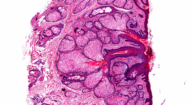
Answer of Dermatopathology Case 73
 Histochemistry : Methenamine silver stain shows Histoplasma capsulatum fungi.
Visit: Dermatopathology site
Visit : Pathology of Histoplasmosis
Abstract:
Histoplasmosis Presenting as a Cutaneous Malignancy of the Eyelid. Ophthal Plast Reconstr Surg. 2010 Sep 20.
Cutaneous histoplasmosis is an uncommon infection and can occur as a primary infection. A manifestation imitating a cutaneous neoplasm is rare, and eyelid involvement is rarer still. The authors report a case of histoplasmosis that presented as an ulcerated lesion on the lower eyelid margin that clinically resembled a basal cell carcinoma. Given its worldwide distribution, it is important to include this disease in the differential diagnosis of nonhealing eyelid lesions. Biopsy and tissue culture are paramount to establishing the diagnosis. This case describes a rare presentation of histoplasmosis on the eyelid and highlights the importance of histopathologic evaluation.
Primary Cutaneous Histoplasmosis in a HIV-Positive Individual.J Glob Infect Dis.2010 May;2(2):112-5.
A 31-year-old human immunodeficiency virus-positive male who presented to the Dermatology Outpatient Department with complaints of red, raised lesions on the face of 2 weeks duration was, on examination, found to have multiple papulonodular lesions on the face with associated cervical and axillary lymphadenopathy. There was history of local injury on the face 6 months prior to the development of symptoms. Skin biopsy revealed multiple round to oval spores with surrounding halo intracellularly, confirming the diagnosis of cutaneous histoplasmosis. No systemic involvement was detected on further investigations. The patient responded to oral antifungals in a short duration, confirming the local nature of the presentation. This is probably the first time in the literature that a primary cutaneous manifestation of histoplasmosis is being described in an immunocompromised individual.
Cutaneous histoplasmosis due to Histoplasma capsulatum variety duboisii in an immune competent child. About one case in Abidjan, Côte d'Ivoire. Bull Soc Pathol Exot.2009 Aug;102(3):147-9.
Histoplasmosis is a subcutaneous mycosis caused by dimorphic fungus which is to be found in two types: the capsulatum and duboisii types. The capsulatum type has had an increasing incidence with the HIV-AIDS epidemics but it is not demonstrated that the duboisii one has had the same upward incidence. Signs in children and immunocompetent patient are rarely described during this disease. The diagnosis is often late in the child as it looks like Molluscum contagiosum lesions. We report a case of skin histoplasmosis of duboisii type non associated with HIV infection in a child. Diagnosis has been confirmed by a histopathological test of a nodule biopsy. Medical treatment was successfully based on itraconazol.
Primary cutaneous histoplasmosis: case report on an immunocompetent patient and review of the literature.Rev Soc Bras Med Trop. 2008 Nov-Dec;41(6):680-2.
This report describes a case of primary cutaneous histoplasmosis in a 45-year-old male. The presentation consisted of an erythematous nodule on the back of the right hand, accompanied by nontender regional lymphadenomegaly that developed following local trauma that occurred during military training in a tunnel inhabited by bats. Histological examination of a biopsy specimen from the skin lesion showed granulomatous infiltrate, but did not show fungal elements. Culturing of this material, incubated in Sabouraud agar, showed growth of Histoplasma capsulatum. No evidence of systemic involvement or immunosuppression was found. Treatment with 400 mg/day of itraconazole orally for six months resulted in complete remission of the lesion, which was maintained one year after the end of the treatment.


Comments
Post a Comment