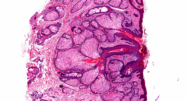
Answer of Dermatopathology Case 75
Granuloma Faciale
Visit: Dermatopathology site
Abstract:
Granuloma faciale with disseminated extra facial lesions. Dermatol Online J.2010 Jun 15;16(6):5.
Granuloma faciale (GF) is a rare cutaneous disorder categorized as a localized form of small vessel vasculitis. Clinically, it manifests as single or multiple, well-demarcated, red-brown plaques, papules and nodules, nearly always confined to the face. Herein, we report a 39-year-old man with multiple red-brown, infiltrated plaques on his face and extrafacial lesions on the back, shoulders, and both arms. Skin biopsy revealed typical histopathological findings of GF. The patient failed to respond to pulsed dye laser, but intralesional triamcinolone combined with cryotherapy led to an acceptable response.
Granuloma faciale: Case report and review. Dermatol Online J. 2009 Dec 15;15(12):3.
Granuloma faciale (GF) is a rare benign chronic inflammatory dermatosis usually appearing only on the face. The lesions of GF typically present as single, asymptomatic, erythematous, non-changing nodules or plaques. We present an illustrative case of GF and briefly review available treatment options.
Granuloma faciale. Pathologica. 2007 Oct;99(5):306-8.
Granuloma faciale is a rare, benign skin condition that usually occurs on the face. Using an exemplary case of granuloma faciale, we will present the clinical and histological characteristics of this dermatosis. A 49-year-old man presented with a 6-month history of a 10 mm-diameter asymptomatic papulo-nodular red-brown lesion of the nose. A biopsy specimen led to the diagnosis of granuloma faciale. The patient received a session of pulsed-dye laser therapy, which led to significant improvement. This benign and usually isolated dermatosis can more rarely be extrafacial. It may often be mistaken for other benign dermatoses (sarcoidosis, discoid lupus erythematosus) as well as for malignant dermatoses (lymphoma, basal cell carcinoma).


Comments
Post a Comment