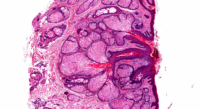
Answer of Dermatopathology Case 79
Alopecia Areata
Visit: Dermatopathology site
Abstract:
'Follicular Swiss cheese' pattern--another histopathologic clue to alopecia areata.J Cutan Pathol. 2011 Feb;38(2):185-9.
Yellow dots are the most useful dermoscopic criterion in the clinical diagnosis of alopecia areata and correspond histopathologically with dilated follicular infundibula. They are found in about 95% of alopecia areata cases and help to differentiate alopecia areata from trichotillomania, telogen effluvium and from scarring alopecias. Histopathology of alopecia areata differs with disease activity and dermatopathologist, therefore, heavily depends on other diagnostic features. Objective of the study was to determine the frequency of dilated follicular infundibula, peribulbar lymphocytic infiltrate, inflammatory infiltrates of lymphocytes and eosinophils within fibrous streamers and a shift to catagen/telogen follicles in alopecia areata. Histopathologic features of 56 specimens of 33 patients were correlated with clinical findings and alopecia areata subtype. Results: 57% of all biopsies showed dilated follicular infundibula, regardless of horizontal or vertical sectioning of the slides. Dilated follicular infundibula showed a maximum occurrence of 66% in the recovery stage of alopecia areata and were seen in 33% of alopecia areata incognita. In conclusion, dilated follicular infundibula, reminiscent of a Swiss cheese in horizontally sectioned slides, is an exceedingly useful criterion in the histopathologic diagnosis of alopecia areata and are of great help in the daily routine to recognize alopecia areata.
Histopathologic features of alopecia areata: a new look. Arch Dermatol. 2003 Dec;139(12):1555-9.
BACKGROUND: A peribulbar lymphocytic infiltrate is the expected histologic feature of alopecia areata, but it is absent in many scalp biopsy specimens. Other diagnostic criteria are needed.
OBJECTIVE: To establish the histologic features of alopecia areata in scalp biopsy specimens taken from different types of alopecia areata, using follicular counts to relate biopsy findings to stages of the disease.
METHODS: Fifty consecutive new patients with alopecia areata were studied. Four-millimeter punch biopsy specimens were taken from the scalp in areas of recent, active hair loss; old, inactive hair loss; or recent hair regrowth. Specimens were sectioned horizontally. Terminal and vellus-like hairs were counted. Inflammation and fibrosis around lower and upper follicles were rated.
RESULTS: The histopathologic features of alopecia areata were not significantly affected by the sex, age, and race of the patient or by the type, percentage of hair loss, total duration, or regression of alopecia areata. The major factor affecting the histopathologic features was the duration of the current episode of alopecia areata. In the acute stage, bulbar lymphocytes surrounded terminal hairs in early episodes and miniaturized hairs in repeated episodes. In the subacute stage, decreased anagen and increased catagen and telogen hairs were characteristic. In the chronic stage, decreased terminal and increased miniaturized hairs were found, with variable inflammation. During recovery, increasing numbers of terminal anagen hairs from regrowth of miniaturized hairs and a lack of inflammation were noted.
CONCLUSIONS: The histopathologic features of alopecia areata depend on the stage of the current episode. Alopecia areata should be suspected when high percentages of telogen hairs or miniaturized hairs are present, even in the absence of a peribulbar lymphocytic infiltrate.
Alopecia areata update: part I. Clinical picture, histopathology, and pathogenesis. J Am Acad Dermatol. 2010 Feb;62(2):177-88, quiz 189-90.
Alopecia areata (AA) is an autoimmune disease that presents as nonscarring hair loss, although the exact pathogenesis of the disease remains to be clarified. Disease prevalence rates from 0.1% to 0.2% have been estimated for the United States. AA can affect any hair-bearing area. It often presents as well demarcated patches of nonscarring alopecia on skin of overtly normal appearance. Recently, newer clinical variants have been described. The presence of AA is associated with a higher frequency of other autoimmune diseases. Controversially, there may also be increased psychiatric morbidity in patients with AA. Although some AA features are known poor prognostic signs, the course of the disease is unpredictable and the response to treatment can be variable. Part one of this two-part series on AA describes the clinical presentation and the associated histopathologic picture. It also proposes a hypothesis for AA development based on the most recent knowledge of disease pathogenesis. LEARNING OBJECTIVES: After completing this learning activity, participants should be familiar with the most recent advances in AA pathogenesis, recognize the rare and recently described variants of AA, and be able to distinguish between different histopathologic stages of AA.


Comments
Post a Comment