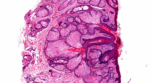
Answer of Dermatopathology Case 100
Retiform Hemangioendothelioma
Visit: Dermatopathology Site
Visit: Pathology of Hemangioendothelioma
Abstract:
Retiform haemangioendothelioma: a case report.Ann Pathol. 2009;29(6):491-4.
Retiform haemangioendothelioma is a locally aggressive, very rarely metastasizing vascular lesion. Histologically, it is characterized by distinctive arborizing blood vessels resembling "rete testis" and lined by endothelial cells with characteristic hobnail morphology. We present an additional case, in the leg of a 64-year-old patient. We discuss the classification of hemangioendotheliomas. The term hemangioendothelioma should be restricted to vascular tumours of "intermediate malignancy" but has been used to designate tumours with variable histological features and clinical behaviour. Spindle cell hemangio(endothelio)ma is currently regarded as a benign reactive lesion. Kaposiform hemangioendothelioma is potentially lethal due to consumption coagulopathy but no metastasizing case has been reported. Epithelioid hemangioendothelioma is associated with a significant metastatic risk and has been included in the category of malignant vascular tumors. The vascular lesions fulfilling the strict definition of hemangioendothelioma include retiform hemangioendothelioma, papillary intralymphatic
angioendothelioma"Dabska's tumor",composite hemangioendothelioma and perhaps the controversial polymorphic hemangioendothelioma.
A case of retiform-hemangioendothelioma with unusual presentation and aggressive clinical features.Int J Clin Exp Pathol. 2010 May 12;3(5):528-33.
Retiform hemangioendothelioma (RH) is an extremely rare low-grade angiosarcoma mainly involving the skin and subcutaneous tissue. Clinically patients often present with an asymptomatic slow-growing solitary nodular or plaque-like lesion. RH is characterized by frequent local recurrences but a very low metastatic rate. Here we reported a case of RH in a 61-year-old Chinese woman who presented with a rapid growing cutaneous plaque-like lesion on her right scalp, followed by another lesion behind the right ear. The lesions were associated with paroxysmal sharp needle-stabbing like headache. She underwent wide excision and skin engraftment. Three months post surgery, she experienced tumor recurrence, and died 9 months after the initial diagnosis.
Retiform hemangioendothelioma developed on the site of an earlier cystic lymphangioma in a six-year-old girl. Am J Dermatopathol. 2011 Oct;33(7):e84-7.
Retiform hemangioendothelioma (RH) is a rare low-grade malignancy angiosarcoma, with a high rate of local recurrence and a low metastatic risk. A 6 year-old girl with a large cervical cystic lymphangioma diagnosed by ultrasound and Doppler ultrasound, which showed a large multiloculated anechoic cyst with no flow. The lymphangioma was treated with injections of Picibanil (OK-432). The tumor regressed, but after a year, she developed a poorly limited infiltrated plaque spreading out regularly over her chest, back, and shoulder. The biopsy showed a poorly limited dermal and subcutaneous vascular proliferation composed of elongated arborising vessels lined with ovoid endothelial cells in a hobnail pattern. In addition, the deep part of the lesion showed typical features of a papillary intralymphatic angioendothelioma pattern (PILA) or Dabska tumor. The endothelial cells strongly expressed podoplanin (D2-40). A diagnosis of RH with focal areas of PILA was reached. The girl died 8 months after surgery of hypovolemic shock in a context of diffuse lymphangiomatosis with pulmonary localization. To our knowledge, RH has hardly ever been described in children. This entity exhibits a continuum with the PILA, sharing not only morphological and immunohistochemical similarities but also its ability to develop in a context of a vascular anomaly, particularly a lymphangioma. The role of Picibanil in the development of this tumor can be discussed.
A rare angiosarcoma: retiform haemangioendothelioma.J Laryngol Otol. 2011 Sep 5:1-3.
Objective:We report the case of a rare angiosarcoma, retiform haemangioendothelioma, in an 18-year-old young man, which presented as a recurrent ulcerating lesion of the left pinna.
Method:Case report and literature review of retiform haemangioendothelioma. This is a low grade angiosarcoma with a high local recurrence rate and low metastasis rate, and was first described in 1994 by Calonje et al.
Results: This patient represents only the third report of lymph node metastasis in a case of retiform haemangioendothelioma. To date, 31 cases of the tumour have been reported. Histological diagnosis of this group of vascular neoplasms can be challenging, as their histopathological appearance is intermediate between haemangioma and angiosarcoma. Conclusion:Surgical excision remains the primary treatment modality, with adjuvant radiotherapy recommended in patients with large tumour size, local recurrence and lymph node metastasis, as seen in this case.
Retiform hemangioendothelioma: presentation of a case expressing D2-40.J Cutan Pathol. 2009 Sep;36(9):987-90.
Retiform hemangioendothelioma (RH) is a low-grade angiosarcoma with low metastatic risk, usually occurring as a single lesion on the trunk or extremity in middle-aged adults.
Histopathology shows a distinctive pattern with arborizing blood vessels arranged in a retiform pattern (similar to rete testis tissue) and focal papillae with fibrosclerotic (hyaline) cores. The blood vessels are lined by comparatively monomorphic endothelial cells, frequently presenting a hobnail pattern. We report a case of RH presenting as an indolent brownish plaque on the back of a 17-year-old male. Surgical resection and sentinel lymph node biopsy showed no evidence of metastasis. In contrast to the recent literature, this case of RH showed positivity for D2-40, a marker of lymphatic endothelium. We also report ultrastructural findings for this case of RH.


Comments
Post a Comment